STAY REPORT
今中隊員の南極滞在記
3.南極の生物編
株式会社アイティー技研 代表取締役の今中です。
私は2004年11月~2005年3月の約4ヵ月間、第46次南極地域観測隊として
南極で微生物の探索業務を行いました。
その南極滞在記を4回に分けてご紹介いたします。
このページでは、南極滞在記その3「南極の生物編」をお楽しみください。
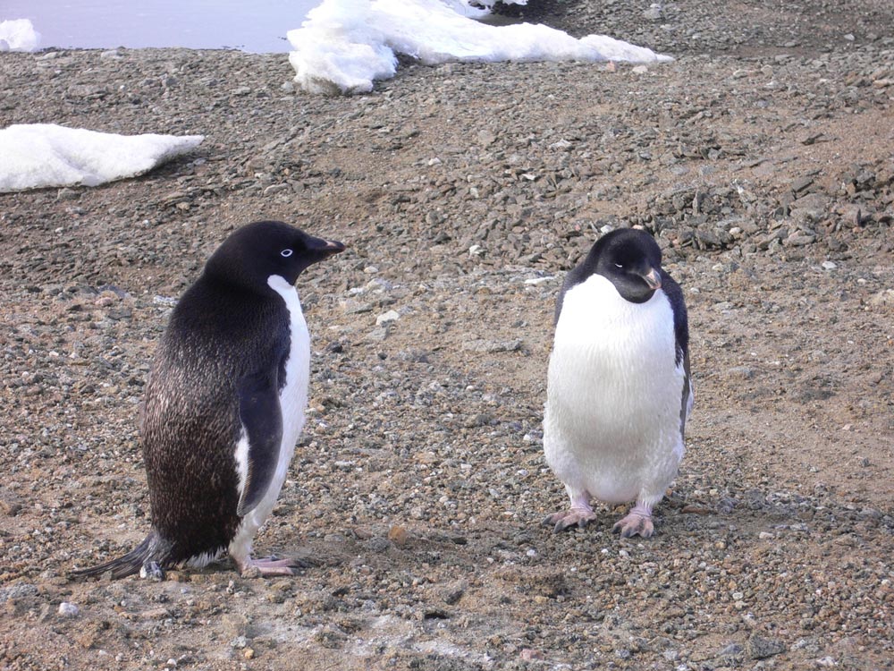
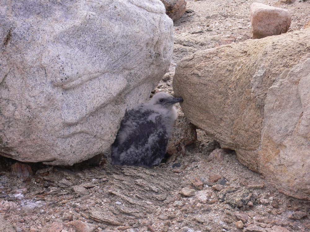
ペンギンの体は保護色になっています。空から見ると黒く、海の下から見ると白っぽく見えるようになっています。
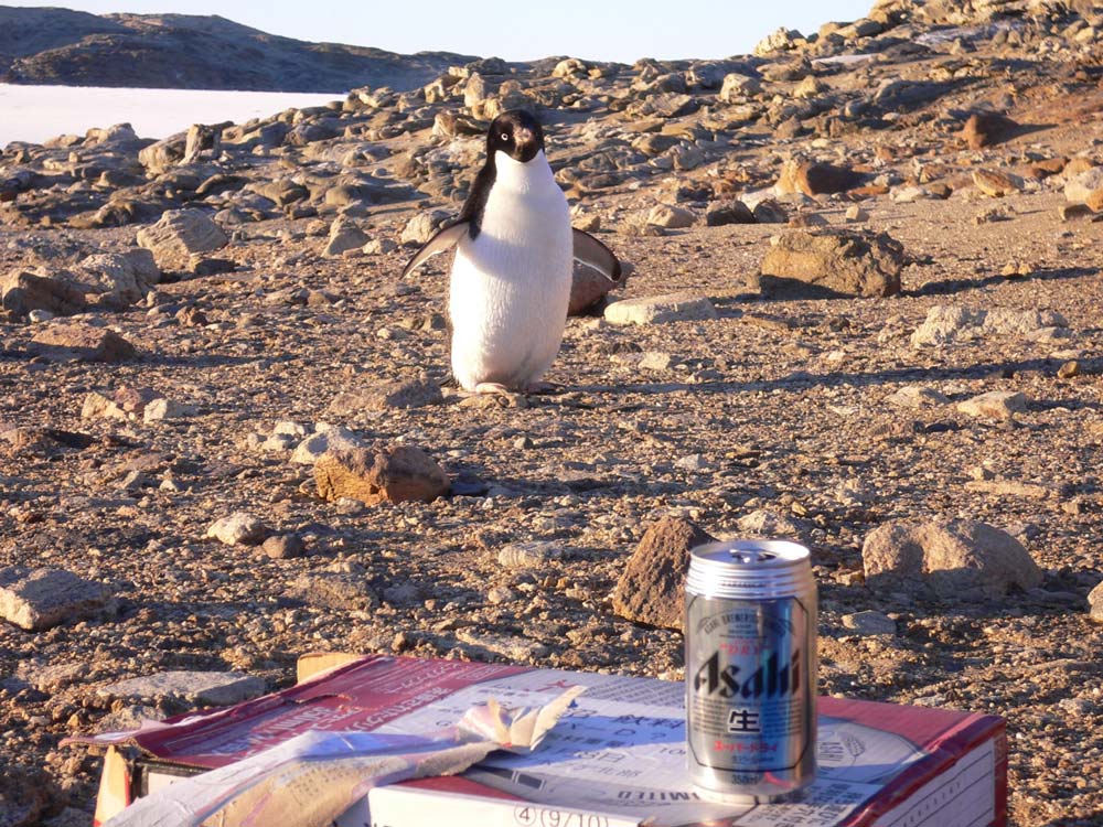
仕事終わりにビールを飲んでいたら、ペンギンが寄ってきました。

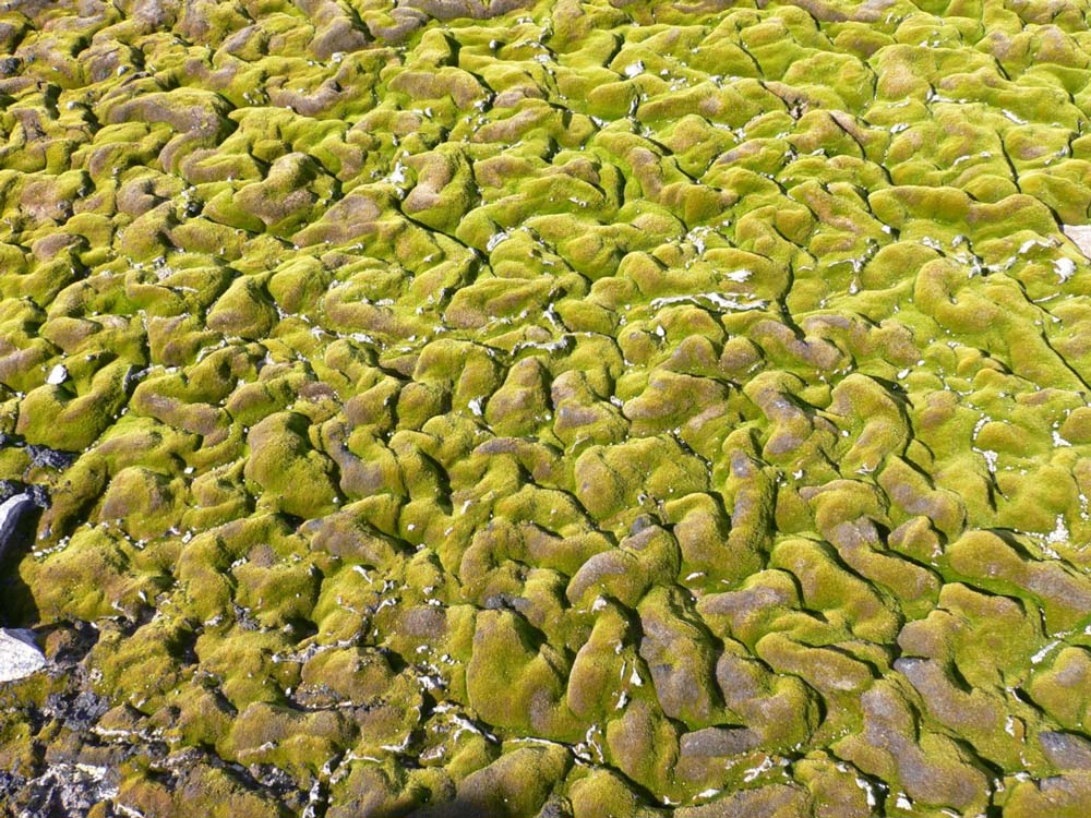
コケの絨毯
コケ:紫外線が強いので、表面は死に青い部分で生長している
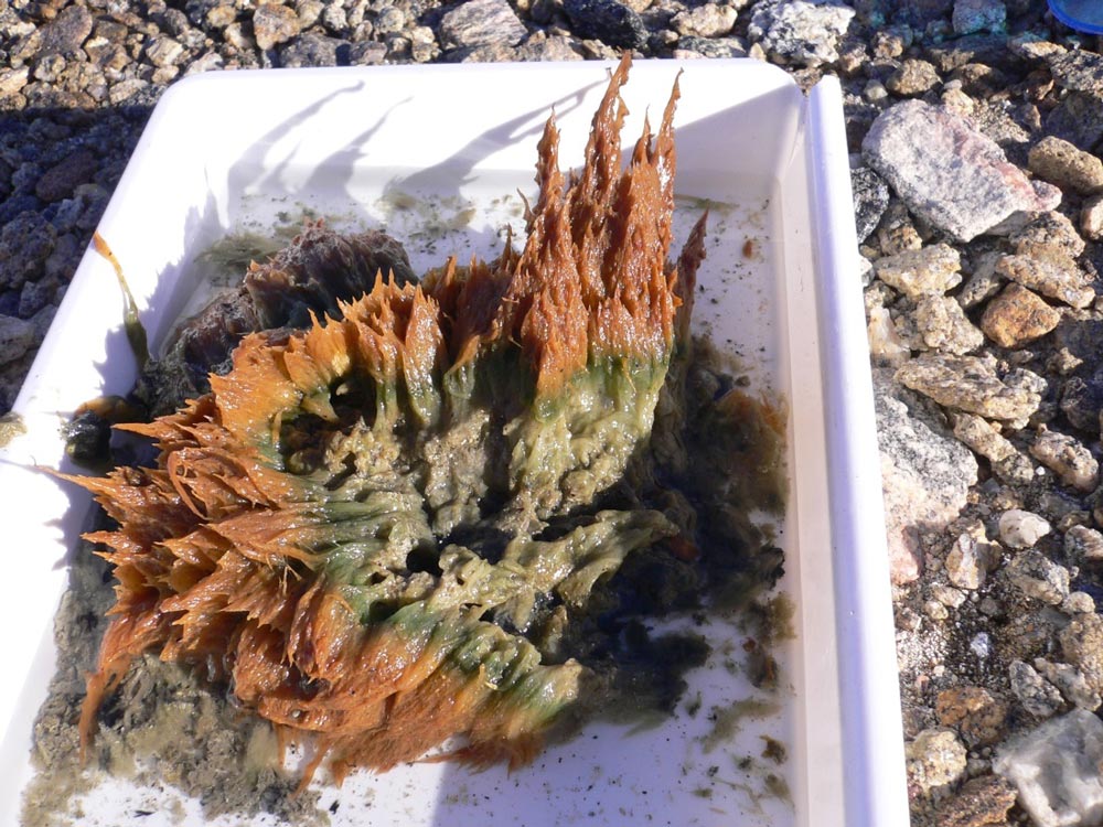
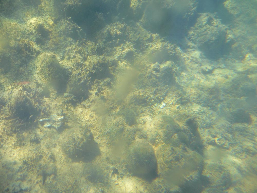
藻類:青い部分で生長している
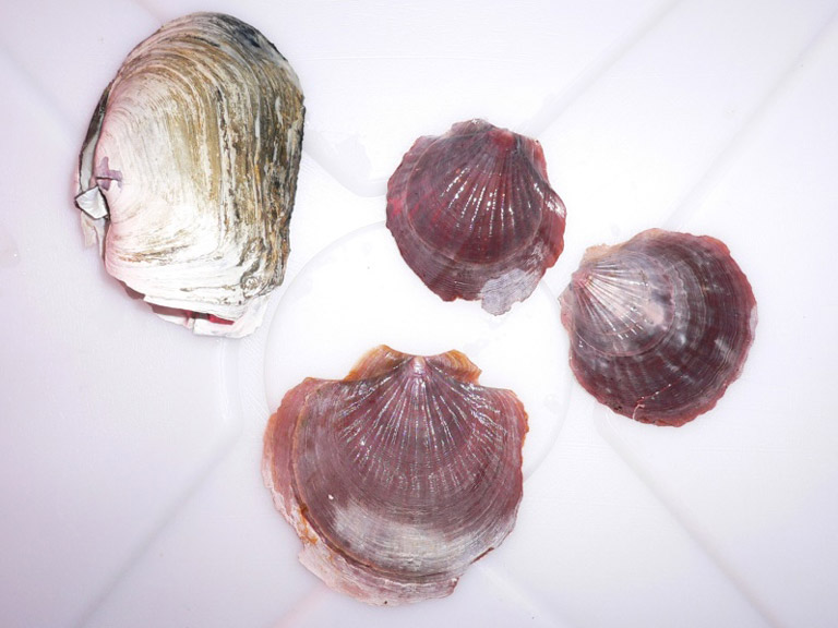
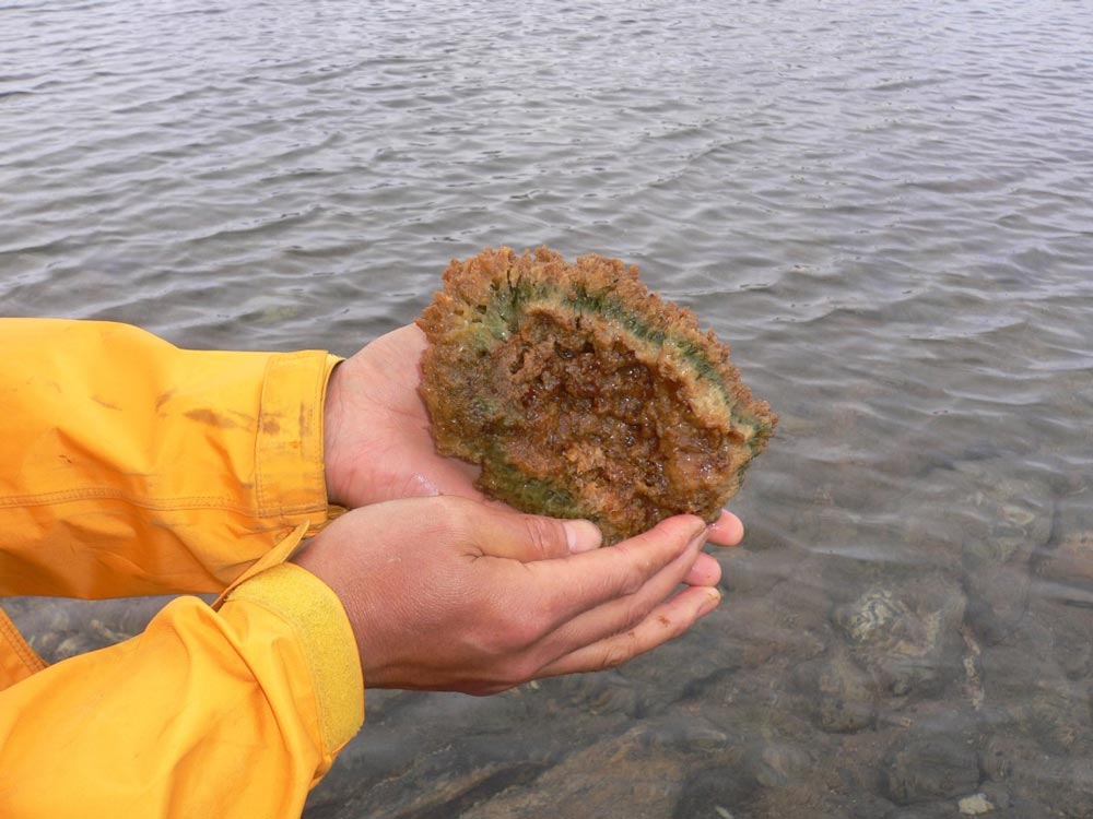
コケ坊主
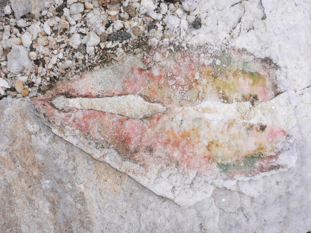
岩を半折したところ
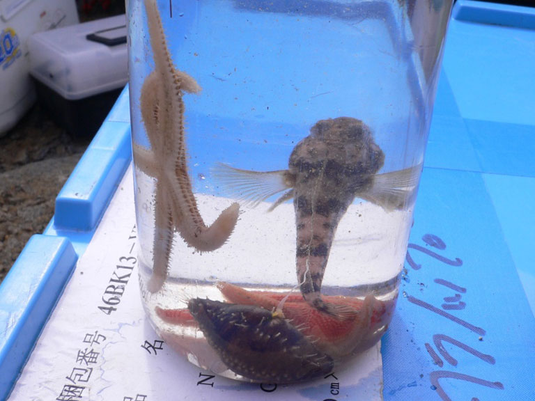
白い岩の隙間に微生物の共生が見られます。
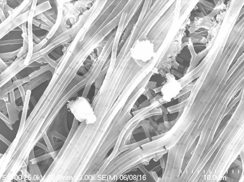
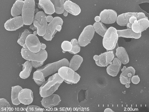
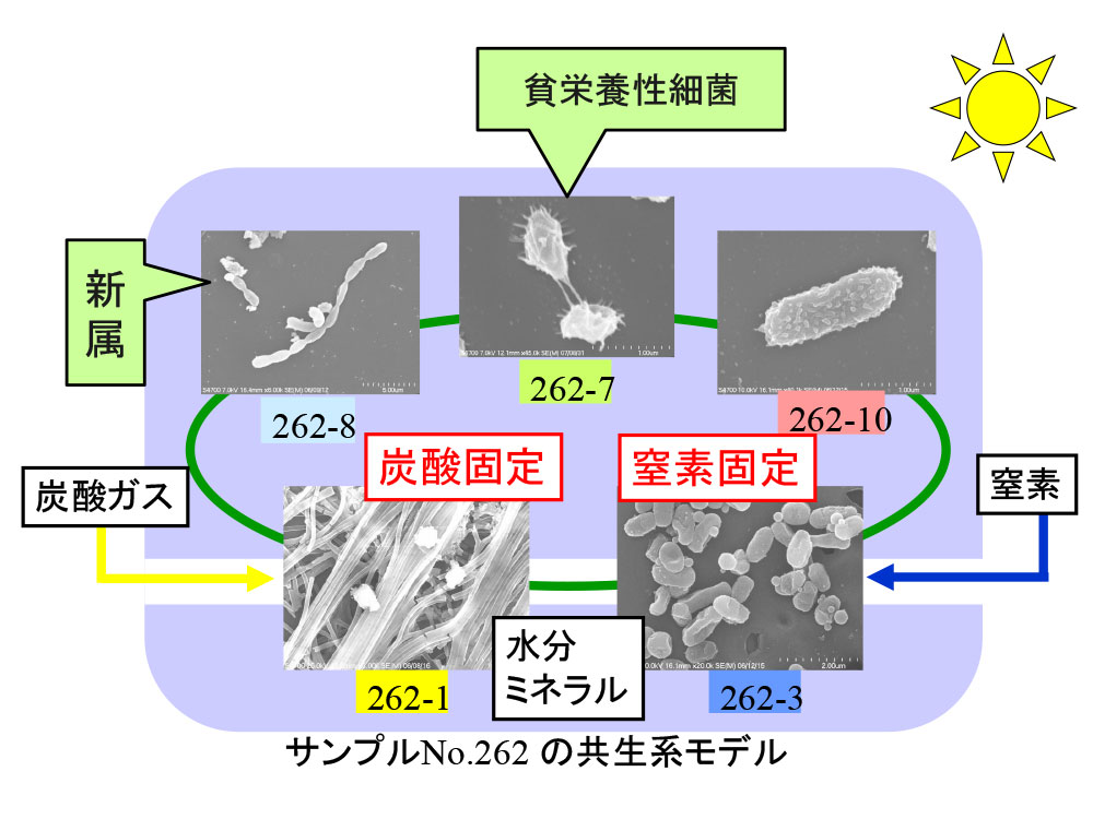
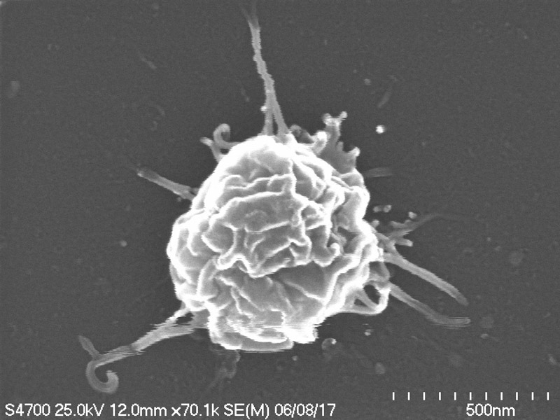
南極から分離した新属の貧栄養菌です。細胞から突起物が出ているのが特徴です。
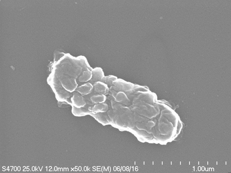
身体のあちこちから細胞が分裂しています。
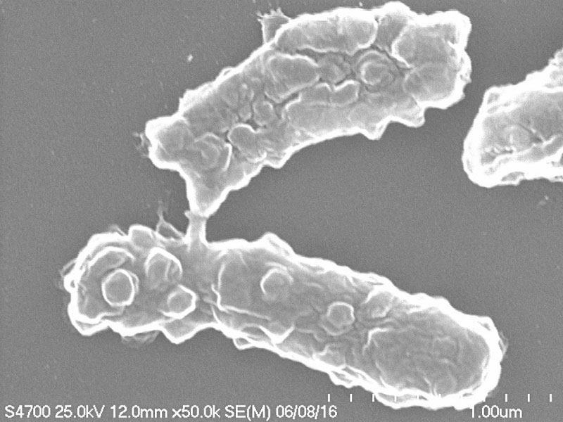
細胞分裂が不自由なため、枝豆状になっています。
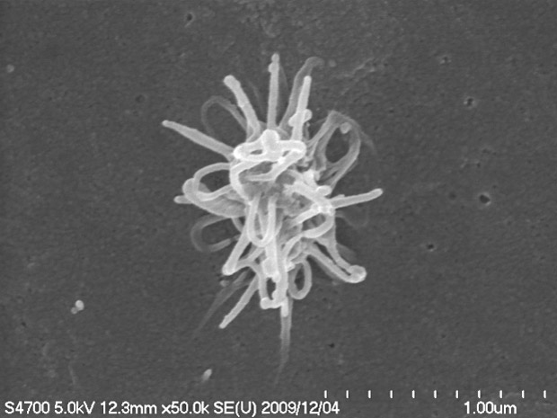
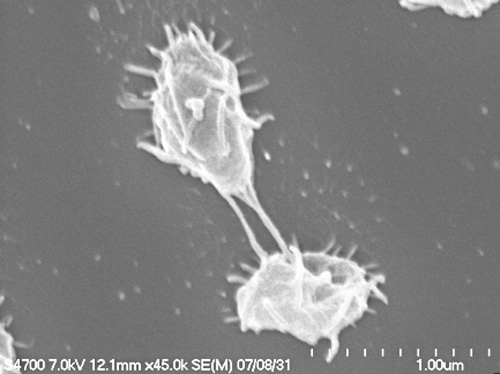
多形性を示しています。1つの細胞からできており、千手観音のようです。
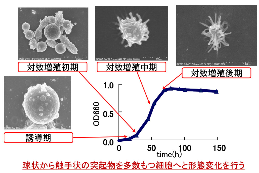
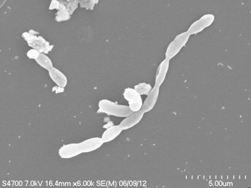
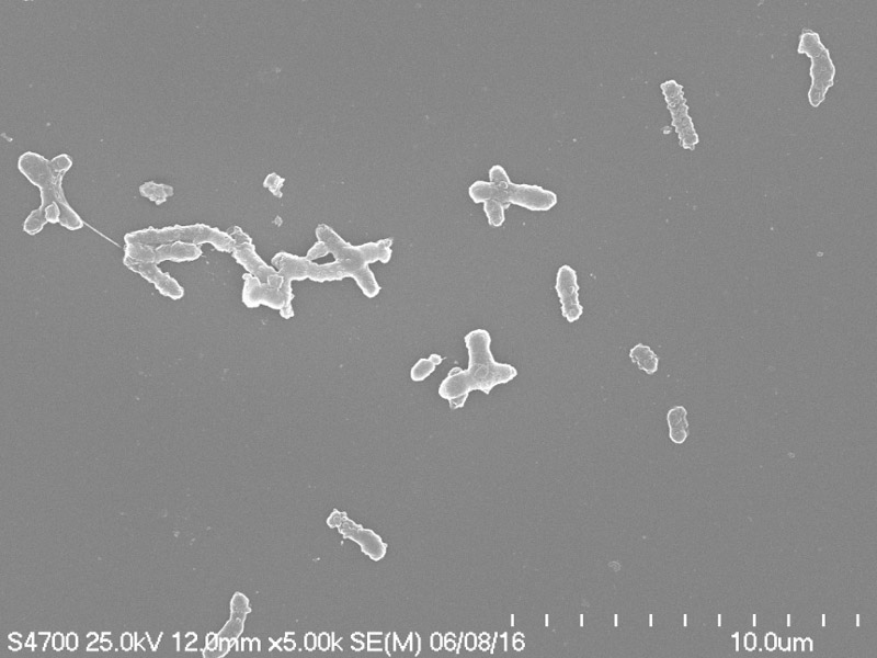
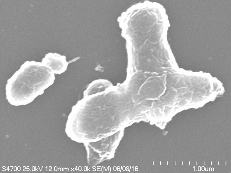
微生物の形態観察です。形態観察は走査型電子顕微鏡(SEM)で行いました。 262-7株は液体培地で生育させた菌体と固体培地で生育させた菌体は異なる特徴を持っていることが分かりました。 液体培養では262-7株は誘導期に球状で、対数増殖初期に細胞から突起物が見え始め、 対数増殖中期でさらに触手状突起物は伸び、対数増殖後期で細胞の短軸の長さにまで伸びていました。 このように、球状から触手状突起物を多数もつ細胞へと形態変化を行うことが分かりました。
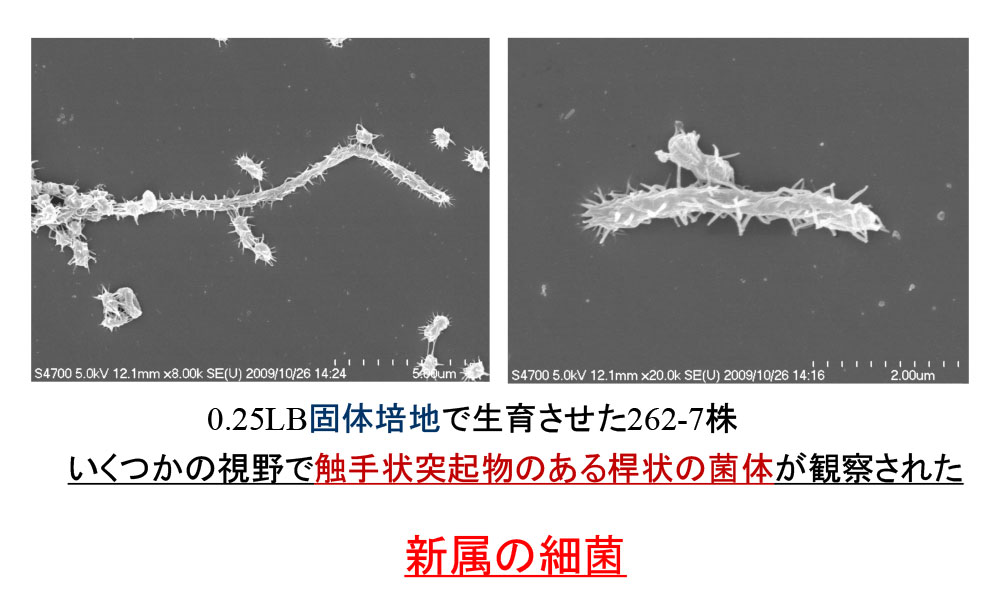
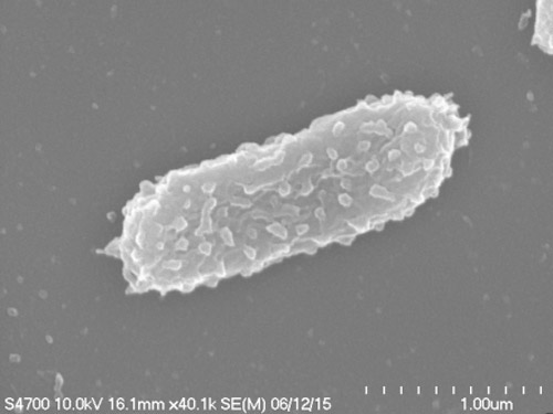
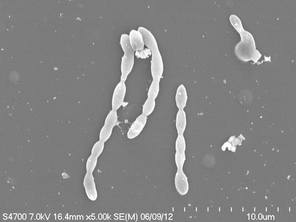
こちらは固体培地上の細胞のSEM観察画像です。 固体培養ではいくつかの視野で触手状の突起物のある桿状菌体が観察されました。 このように細長くなる原因として、不完全な細胞分裂が行われている可能性が考えられました。 そこでこの菌の核酸染色し、核酸の局在の様子を観察することにしました。 棘は栄養を吸収しやすくするのではと考えています。
続いて、南極滞在記その4
「南極の大自然編」
をお楽しみください!